- Obtenez des conseils d’acquisition étape par étape pour simplifier l’imagerie 3D
-
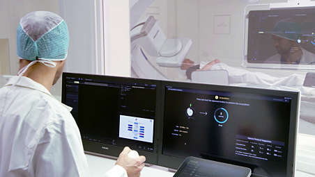
Obtenez des conseils d’acquisition étape par étape pour simplifier l’imagerie 3D
Pour permettre aux utilisateurs d’acquérir des images 3D de grande qualité, quel que soit leur niveau d’expérience [1], SmartCT Vaso fournit un guidage étape par étape et des aides visuelles pendant l’acquisition. Cela inclut la configuration de la salle, l’isocentrage de dose nulle, ainsi que la suggestion d’une injection de produit de contraste adaptée pour le protocole sélectionné. - Inspection directe des images avec visualisation 3D avancée depuis la table
-
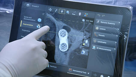
Inspection directe des images avec visualisation 3D avancée depuis la table
Une fois acquise, l’image 3D s’affiche automatiquement sur le module à écran tactile dans le mode de rendu correspondant pour un examen direct depuis la table. Vous pouvez faire défiler, zoomer, réaliser un panoramique et faire pivoter l’image, régler l’épaisseur de coupe et utiliser la reconstruction multiplanaire sur l’écran tactile. - Vérification du positionnement des stents du flow diverter pendant une procédure
-
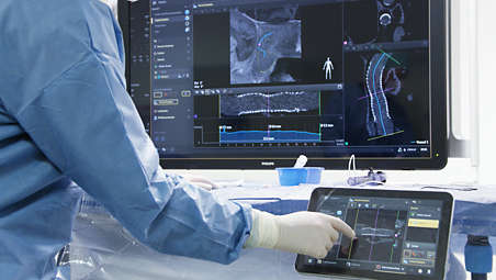
Vérification du positionnement des stents du flow diverter pendant une procédure
La technique SmartCT Vaso est de plus en plus utilisée pour le suivi des anévrismes traités avec des stents de flow diverter afin de vérifier le positionnement du dispositif. Il offre des images de grande qualité pour évaluer les flow diverters jusqu’à la veine perforante. SmartCT Vessel Analysis permet une analyse du vaisseau et une vérification du positionnement du dispositif grâce à des vues informatisées redressées, convexes et transversales.
Obtenez des conseils d’acquisition étape par étape pour simplifier l’imagerie 3D
Obtenez des conseils d’acquisition étape par étape pour simplifier l’imagerie 3D
Obtenez des conseils d’acquisition étape par étape pour simplifier l’imagerie 3D
Inspection directe des images avec visualisation 3D avancée depuis la table
Inspection directe des images avec visualisation 3D avancée depuis la table
Inspection directe des images avec visualisation 3D avancée depuis la table
Vérification du positionnement des stents du flow diverter pendant une procédure
Vérification du positionnement des stents du flow diverter pendant une procédure
Vérification du positionnement des stents du flow diverter pendant une procédure
- Obtenez des conseils d’acquisition étape par étape pour simplifier l’imagerie 3D
- Inspection directe des images avec visualisation 3D avancée depuis la table
- Vérification du positionnement des stents du flow diverter pendant une procédure
- Obtenez des conseils d’acquisition étape par étape pour simplifier l’imagerie 3D
-
Obtenez des conseils d’acquisition étape par étape pour simplifier l’imagerie 3D
Pour permettre aux utilisateurs d’acquérir des images 3D de grande qualité, quel que soit leur niveau d’expérience [1], SmartCT Vaso fournit un guidage étape par étape et des aides visuelles pendant l’acquisition. Cela inclut la configuration de la salle, l’isocentrage de dose nulle, ainsi que la suggestion d’une injection de produit de contraste adaptée pour le protocole sélectionné. - Inspection directe des images avec visualisation 3D avancée depuis la table
-
Inspection directe des images avec visualisation 3D avancée depuis la table
Une fois acquise, l’image 3D s’affiche automatiquement sur le module à écran tactile dans le mode de rendu correspondant pour un examen direct depuis la table. Vous pouvez faire défiler, zoomer, réaliser un panoramique et faire pivoter l’image, régler l’épaisseur de coupe et utiliser la reconstruction multiplanaire sur l’écran tactile. - Vérification du positionnement des stents du flow diverter pendant une procédure
-
Vérification du positionnement des stents du flow diverter pendant une procédure
La technique SmartCT Vaso est de plus en plus utilisée pour le suivi des anévrismes traités avec des stents de flow diverter afin de vérifier le positionnement du dispositif. Il offre des images de grande qualité pour évaluer les flow diverters jusqu’à la veine perforante. SmartCT Vessel Analysis permet une analyse du vaisseau et une vérification du positionnement du dispositif grâce à des vues informatisées redressées, convexes et transversales.
Obtenez des conseils d’acquisition étape par étape pour simplifier l’imagerie 3D
Obtenez des conseils d’acquisition étape par étape pour simplifier l’imagerie 3D
Obtenez des conseils d’acquisition étape par étape pour simplifier l’imagerie 3D
Inspection directe des images avec visualisation 3D avancée depuis la table
Inspection directe des images avec visualisation 3D avancée depuis la table
Inspection directe des images avec visualisation 3D avancée depuis la table
Vérification du positionnement des stents du flow diverter pendant une procédure
Vérification du positionnement des stents du flow diverter pendant une procédure
Vérification du positionnement des stents du flow diverter pendant une procédure
Produits associés
Alternative products
-
Azurion 7 M20
- Système d’imagerie interventionnelle monoplan fixé au sol/plafond avec capteur plan 20“
- Améliorez la visibilité pour diverses procédures vasculaires, oncologiques et cardiaques avec une excellente qualité d’image
- Contrôlez toutes les applications pertinentes depuis la table via le module à écran tactile central
Voir le produit
-
Azurion 7 B20/15
- Système d’imagerie interventionnelle biplan avec un capteur plan 20“ frontal et un capteur plan 15“ latéral
- Renforce la sécurité pendant les interventions neurologiques telles que l’AVC ischémique et le traitement de l’anévrisme cérébral
- Rend entièrement visibles les détails de malformations complexes ainsi que les flow diverters les moins radio-opaques
- Faites l’expérience de processus cliniques simples et fluides avec nos caractéristiques dédiées dans le domaine de la neurologie
Voir le produit
-
Azurion 7 M20 with FlexArm
- Système d’imagerie interventionnelle monoplan fixé au plafond avec un capteur plat 20”
- Pivote sur pas moins de huit axes pour créer une flexibilité quasiment illimitée pour l’imagerie
- Le système de contrôle intelligent des mouvements Axsys permet une commande plus indépendante pour les médecins
- Contrôlez toutes les applications pertinentes depuis la table via le module à écran tactile central
Voir le produit


-
Azurion 7 M20
Découvrez un nouveau monde de performances cardiaques et vasculaires interventionnelles avancées sur la série Azurion 7 avec capteur plan 20”. Cette solution de radiologie interventionnelle de pointe permet d’offrir une prise en charge de haute qualité à vos patients et d’améliorer votre efficacité opérationnelle en associant excellence clinique et innovation des processus. Contrôlez facilement toutes les applications pertinentes à partir d’un seul écran tactile depuis la table, afin de prendre des décisions rapides et éclairées dans l’environnement stérile.
Voir le produit
-
Azurion 7 B20/15
Le système Philips Azurion vous permet d’effectuer une large gamme de procédures interventionnelles de routine comme complexes avec simplicité et fiabilité, au travers d’une expérience utilisateur unique. Des fonctionnalités avancées intégrées à une géométrie de système innovante permettent d’améliorer le processus de travail, ce qui vous aide à optimiser les performances de vos équipements et améliorer la prise en charge de vos patients.
Voir le produit
-
Azurion 7 M20 with FlexArm
Ce système avec suspension plafonnière offre une flexibilité d’imagerie illimitée pour un grand nombre de procédures, ainsi qu’une grande liberté de positionnement pour les équipes médicales. Sa configuration compacte offre un environnement très rentable, prêt pour les procédures de demain. Contrôlez facilement toutes les applications pertinentes à partir d’un seul écran tactile depuis la table, afin de prendre des décisions rapides et éclairées dans l’environnement stérile. Parce qu’il travaille autour de vous, le système Philips Azurion équipé d’un statif FlexArm vous aide à optimiser les performances de votre salle interventionnelle et à prodiguer des soins de meilleure qualité.
Voir le produit
- ¹Nogueira RG, Yoo AJ, Buonanno FS, Hirsch JA. Endovascular approaches to acute stroke, part 2: a comprehensive review of studies and trials. AJNR 2009; 30:859-875